
Research
- Background – why water near a macromolecule’s surface behaves so differently from bulk water, and why that matters for chemistry and biology.
Projects
- Open Instrumentation (New Initiative) – affordable, sensitive open NMR instrumentation as real-time “eyes” for autonomous chemistry.
- Spin Physics for Low-Field NMR – applying our quantitative understanding of ODNP and the DCCT technique to get clean, quantitative signal out of noisy, low-cost, low-field magnets.
- Temperature-Controlled ODNP in Reverse Micelles – reverse micelles as a tunable model system for confined water and protein-surface hydration.
- References – works cited above, numbered as they appear on this page.
Background
Even though water appears uniform on a macroscopic scale, it behaves quite differently on the nanoscale. If we zoom in to the first few layers of water molecules near the surface of a protein or polymer (in fact, near any macromolecule or macromolecular assembly), we see that hydrogen bonds break and individual water molecules move at rates anywhere from two times slower to hundreds of times slower than those in the bulk liquid. We know that these differently behaved “hydration waters” are key to determining interactions.
The future of physical and analytical chemistry will center on the fact that, while tools have been developed for characterizing static structures, we still need techniques that can study dynamic arrangements of molecules and, most urgently, the arrangement of water molecules.
Comprehensive methods for mapping out the structure of the hydration layer do not currently exist. The future of chemistry, broadly, depends on techniques that will map out the structure of the dynamic solvent that surrounds macromolecules. The resulting insight will improve our ability to design drugs and synthetic materials. In particular, by developing technologies that can map out the dynamic structure of hydration water, we unlock an ability to design drugs that bind to proteins currently deemed “undruggable,” we can understand the energetics that cause signaling proteins to bind to their partners, and design polymer materials that conduct water in unique ways.
Most chemists and biologists recognize one very simple example of hydration water: the layer of water surrounding oil molecules that drive the separation of water and oil. But the more general case, especially for macromolecules that present a surface with variable properties to the solvent, is far more interesting and far less understood.
How do proteins alter nearby solvent? Proteins are the molecular motors that drive all life, and most drugs are designed to interact with proteins. It is increasingly clear that water molecules at the surface of proteins control their structure and function; while researchers have developed methods that can study the structure of proteins, there are no accessible methods to spatially map the hydration water. We develop and apply magnetic-resonance-based spectroscopic methods for routine, robust, and accessible measurements of the properties of these key hydration water molecules.
A fundamental example. As a fundamental example, consider lipid bilayers – structures that make up cell membranes. Dr. Franck and his colleagues previously developed a specialized technique to characterize water near the bilayer surfaces. Our technique – called Overhauser Effect Dynamic Nuclear Polarization (ODNP) 1,2 – tracks the motion of the water nuclei, and allows us to zoom in on the “hydration water” near the surface of the lipid bilayer (figure below). It allowed them to see that the hydration water moves about 5 times slower than bulk water 3. Furthermore, by dramatically changing the characteristics of the bulk solution, we learned that an order of magnitude change in the viscosity barely affects the diffusion in the hydration layer 3. They have also implemented this technique in systems with membrane proteins 4, DNA 5, and large protein folding chaperones 6. The significant variations in the properties of the hydration water play in important role in how these surfaces interact in nature.
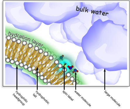
This image illustrates ODNP of water in the hydration layer of a lipid bilayer. This is one example of many studies that are possible with ODNP. A lipid bilayer (the basis for cellular membranes) consists of a self-organized set of molecules with hydrophilic headgroups and hydrophobic tails (see labels above). One can place a spin label (typically a nitroxide group) at a particular location near the surface of the lipid bilayer (via chemical synthesis). A combination of electron- and nuclear-spin resonance (i.e. ODNP) can selectively interrogate the motion of water molecules at a specific location – here inside the hydration layer of the lipid bilayer. For example, employing the setup shown above, we were able to verify that the motion of water molecules within the hydration layer was not sensitive to the presence of a high concentration of large molecules that are not chemically disposed to disrupt the hydration layer. This was even true when the large molecules significantly altered the viscosity of the bulk solution 3.
In the Franck lab, we currently push the limits of the ODNP technique to retrieve new and more detailed information about hydration water, drawing on expertise in spectrometer design and advanced magnetic resonance methods 7,8 that we have already distilled into a cogent, quantitative explanation of the ODNP technique itself 2. That work is organized into three projects, described below.
Open Instrumentation (New Initiative)

Since joining ACERT, we’ve launched a new initiative built around the observation that autonomous chemistry labs need real-time, quantitative feedback on what’s actually happening in a reaction – the “eyes” that let a robot check its own work – and that today’s benchtop NMR instruments aren’t sensitive, flexible, or affordable enough to provide it.
We design inexpensive, customizable, yet sensitive open instrumentation 9,10,11, an approach very much aligned with ACERT’s mission of making advanced magnetic resonance broadly accessible.
Spin Physics for Low-Field NMR
We draw on our expertise in advanced spin physics: applying our quantitative understanding of the Overhauser effect together with the DCCT technique 12, which extracts clean, multiplexed signal from the noisy, inhomogeneous fields typical of low-cost, low-field magnets 13. Together, these let us push sensitive, quantitative magnetic resonance into settings – like an autonomous synthesis robot – where it was previously impractical.
Temperature-Controlled ODNP in Reverse Micelles
We are investigating the water on the insides of reverse micelles, which provide controllable, temperature-tunable pockets of confined water and serve as our model system for confinement effects more broadly 14,15. Atahan Garip is extending this program by developing a fully open and automated temperature-controlled ODNP system, purpose-built for these measurements.
When nanoscale aggregates such as reverse micelles isolate small pockets of water, the resulting confinement alters the hydrogen bonding network; meanwhile, the confinement also slows down the motion of the water molecules. These effects result in a change (respectively) of the electron cloud density and of the lifetime of the spin states, and magnetic resonance can measure both of these. We have developed a measurement that reads out the correlation between the hydrogen bonding strength and the rotational motion in confined water pools. By measuring this correlation as a function of the size of the water pool, we’ve identified a distinct inflection point at a couple nanometers at which the water matrix changes dramatically.
We also continue to map out more subtle details in the water along the surface of proteins, both globular signaling proteins as well as trans-membrane proteins, employing mutagenesis to attach a spin label at a series of sites on the surface of proteins, including cancer signaling proteins (KRas) as well as transmembrane proton pumps (proteorhodopsin). This allows us to create a set of samples that are primed for measurement of water at a series of different sites, so that we can watch how the translational diffusivity of the water changes as we move from site to site along the surface.
References
See the Publications page for our complete list of publications
- 1. Franck JM, Pavlova A, Scott JA, Han S. “Quantitative cw Overhauser effect dynamic nuclear polarization for the analysis of local water dynamics.” Prog Nucl Magn Reson Spectrosc. Elsevier B.V.; 2013 Oct;74:33–56. doi:10.1016/j.pnmrs.2013.06.001 PMID:24083461 arXiv:1206.0510
- 2. Franck JMJM, Han S. “Overhauser Dynamic Nuclear Polarization for the Study of Hydration Dynamics, Explained.” In: Wand AJ, editor. Biol NMR Part B. Academic Press; 2019. p. 131–175. doi:10.1016/bs.mie.2018.09.024
- 3. Franck JM, Scott JA, Han S. “Nonlinear Scaling of Surface Water Diffusion with Bulk Water Viscosity of Crowded Solutions.” J Am Chem Soc. 2013 Mar;135(11):4175–4178. doi:10.1021/ja3112912 PMID:23347324
- 4. Hussain S, Franck JM, Han S. “Transmembrane Protein Activation Refined by Site-Specific Hydration Dynamics.” Angew Chemie Int Ed. 2013 Feb;52(7):1953–1958. doi:10.1002/anie.201206147 PMID:23307344
- 5. Franck JM, Ding Y, Stone K, Qin PZ, Han S. “Anomalously Rapid Hydration Water Diffusion Dynamics Near DNA Surfaces.” J Am Chem Soc. 2015 Sept;137(37):12013–12023. doi:10.1021/jacs.5b05813
- 6. Franck JM, Sokolovski M, Kessler N, Matalon E, Gordon-Grossman M, Han S, Goldfarb D, Horovitz A. “Probing Water Density and Dynamics in the Chaperonin GroEL Cavity.” J Am Chem Soc. 2014 July;136(26):9396–403. doi:10.1021/ja503501x PMID:24888581
- 7. Franck JM, Chandrasekaran S, Dzikovski B, Dunnam CR, Freed JH. “Focus: Two-dimensional electron-electron double resonance and molecular motions: The challenge of higher frequencies.” J Chem Phys. 2015;142(21):212302. doi:10.1063/1.4917322
- 8. Franck JM, Demas V, Martin RW, Bouchard L-S, Pines A. “Shimmed matching pulses: Simultaneous control of rf and static gradients for inhomogeneity correction.” J Chem Phys. 2009 Dec;131(23):234506. doi:10.1063/1.3243850 PMID:20025334
- 9. Franck JM, Barnes RP, Keller TJ, Kaufmann T, Han S. “Active cancellation – A means to zero dead-time pulse EPR.” J Magn Reson. 2015 Dec;261:199–204. doi:10.1016/j.jmr.2015.07.005
- 10. Kaufmann T, Keller TJ, Franck JM, Barnes RP, Glaser SJ, Martinis JM, Han S. “DAC-board based X-band EPR spectrometer with arbitrary waveform control.” J Magn Reson. 2013 Oct;235:95–108. doi:10.1016/j.jmr.2013.07.015 PMID:23999530
- 11. Demas V, Franck JM, Bouchard LS, Sakellariou D, Meriles CA, Martin R, Prado PJ, Bussandri A, Reimer JA, Pines A. “‘Ex situ’ magnetic resonance volume imaging.” Chem Phys Lett. 2009 Jan;467(4-6):398–401. doi:10.1016/j.cplett.2008.11.069
- 12. Beaton AA, Guinness A, Franck JM. “A modernized view of coherence pathways applied to magnetic resonance experiments in unstable, inhomogeneous fields.” J Chem Phys. 2022 Nov;157(17):174204. doi:10.1063/5.0105388 arXiv:2202.03313
- 13. Beaton AA, Guinness A, Franck JM. “Rapidly Screening the Correlation between the Rotational Mobility and the Hydrogen Bonding Strength of Confined Water.” J Phys Chem B. 2024 Oct;128(43):10749–10763. doi:10.1021/acs.jpcb.4c05397 PMID:39439388 arXiv:2306.06571
- 14. Beaton AA, Franck JM. “Direct Observation of the Translational Immobilization of Water Molecules Under Nanoscale Confinement.” arXiv; 2023. arXiv:2306.07240
- 15. Shathy R, Guinness A, Franck JM. “Exchange and Microviscosity of Dynamic Nanocompartments.” arXiv; 2024. arXiv:2410.10748